Practical guide
.png)
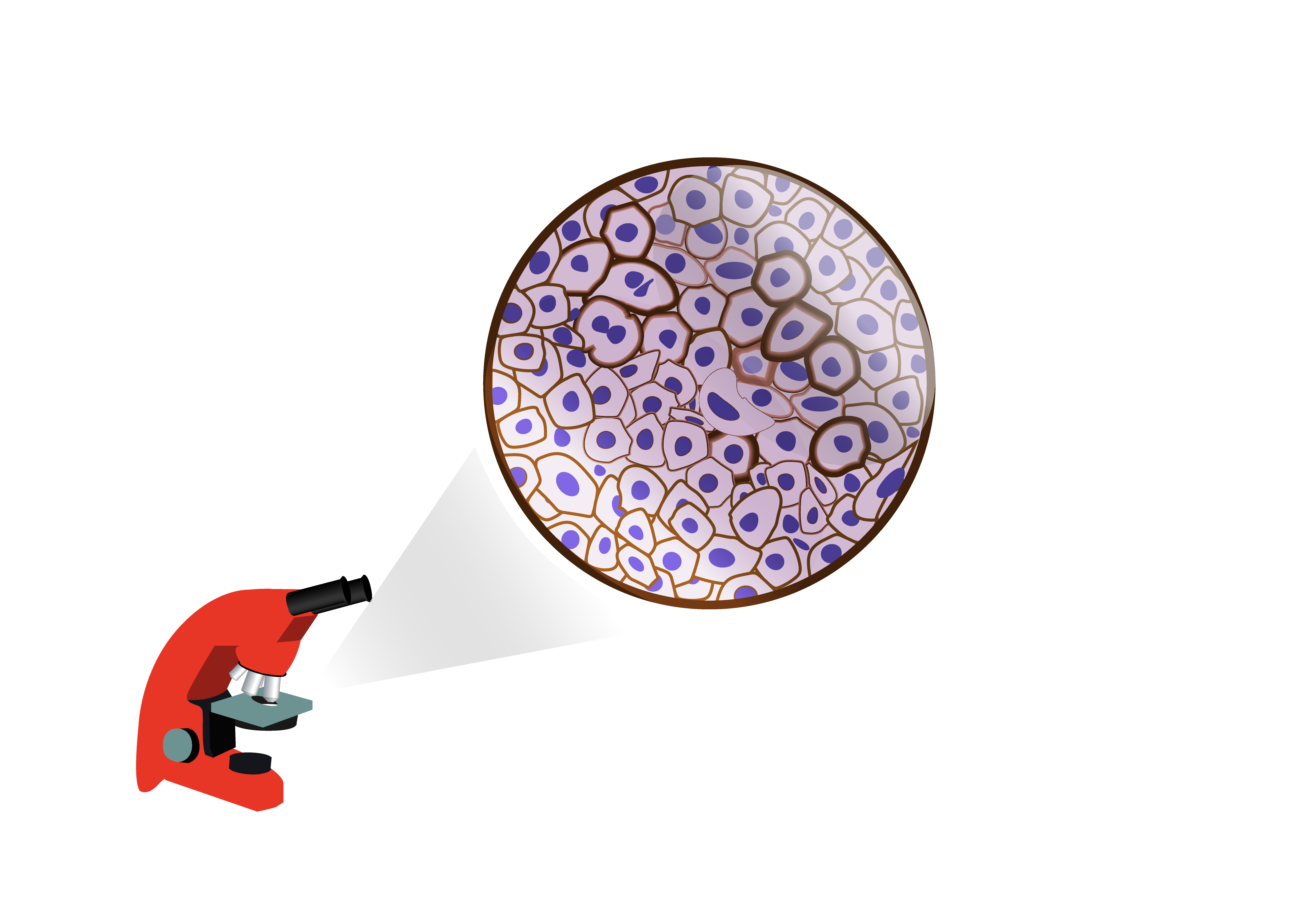
IHC protocol
Immunohistochemistry is a technique that aims to localize a protein in cells within a tissue section by using a specific antibody, which will bind to an antigen of the biological tissue. This technique is widely used, for example, to identify and characterize a tumor (malignancy, classification...).
BAP1, clone C4
BAP1 protein or BRCA1 associated protein-1 (ubiquitin carboxy-terminal hydrolase) is a de-ubiquitination enzyme encoded by BAP1 gene located on human chromosome 3. BAP1 (BRCA1-associated protein 1) binds to the N-terminal domain of BRCA1 and is a tumor suppressor gene.
STAT6, Clone EP325
Solitary fibrous tumors (SFT) are uncommon fibroblastic neoplasms. Often diagnosed by their histological characteristics and CD34 ...
Rat Monoclonal Antibodies - Mouse Specific
Discover our new mouse-specific rat monoclonal antibodies for IHC-P.
Uroplakin II
Uroplakins Ia, Ib, II and III are structural proteins of urothelial cells differentiated. In non-neoplastic urothelial cells, Uroplakins are expressed in the luminal membrane of surface cells.
Cadherin 17
A new marker for gastrointestinal cancers diagnosis.
Cadherin 17 is a non-conventional cadherins and is also referred to as LI-cadherin or CDH17. It is involved in tumor invasion and metastasis...
SOX-10 Antibody
SOX-10 is a very sensitive melanomas marker (97% versus 91% for S-100). It is particularly sensitive for desmoplastic melanomas which are often difficult to detect by other markers of melanocytes. SOX-10 is moderately to strongly expressed in desmoplastic melanomas ...
Arginase 1
Distinction of hepatic metastases from hepatocellular carcinomas (HCC) can present some difficulties. Arginase-1 is a marker recently described in the literature as a new highly specific and sensitive polyclonal antibody for HCC detection...
ALK / p80: Control Cell Lines
9-Core ALK-1 Cell Line and 3-Core Lung Cancer Cell Line Microarray consist of 2 mm diameter deposits of cell lines fixed with formalin and included in paraffin...
MDM2
MDM2 is an important negative regulator of the p53 tumor suppressor. MDM2 is an inhibitor of the P53 transcription, and function like an E3 ubiquitin ligase recognizing the N-terminal domain of trans-activation (TAD) of the tumor suppressor p53. Already known for its oncogene role, it has been shown that MDM2...
SATB2 antibody
SATB2 (Special AT-sequence rich-Binding Protein 2) is a new very specific marker for colorectal cancer (CRC). This protein is selectively expressed in glandular cells of the lower gastrointestinal tract and its expression is conserved in a large majority of primary and metastatic CRCs...
NKX3.1 Antibody
NKX3.1 has been identified as a marker for metastatic tumors. NKX3.1 is positive for prostate staining cells with nuclear staining. Most cases of urothelial carcinoma were found negative for NKX3.1, allowing to distinguish high-grade prostate adenocarcinomas and high-grade infiltrated urothelial carcinomas...
Pan Plus Cytokeratin
Pan Plus Cytokeratin is an excellent alternative to Pan Cytokeratin clone KL-1. This cocktail allows the detection of acid cytokeratins 10, 15, 16 and 19
Cytokeratin KL1 available again !
This antibody recognizes neoplastic and normal cells of epithelial origin but does not recognize cells from the epidermal basal layers.
Its specificity is to recognize cytokeratins 1, 2, 5, 6, 7, 8, 10, 11, 14, 16, 17, 18 and 19.
PAX-8
PAX-8 belongs to the “paired box” transcription factors protein family. This nuclear protein is involved in follicular cells development in the thyroid gland and in the expression of specific thyroid genes. Mutations in PAX-8 gene have been associated with dysgenesis, follicular carcinoma and atypical adenomas of the thyroid gland...
Galectin-3, Clone 9C4
Galectin-3 is a 31 kDa lectin that binds to beta-galactosidase. Galectin-3 is normally expressed in the epithelia of many organs and various inflammatory cells, including macrophages, dendritic cells and Kupffer cells.
Pin cocktail
This antibody cocktail is intended for human P504S (also known as AMACR or α-Methylacyl-CoA-Racemase) and human p63 detection in formalin-fixed paraffin-embedded tissues.
Chemiluminescent Pen - WesternBright ChemiPen
The WesternBright ChemiPen allows you to write or draw on transfer membranes and reveal protein standards, for example.
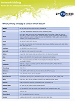
Carcinomas Immunophenotyping using IHC
Find here some scientific information relating the antibodies to be used for specific tissues in diagnosis.
Antibody Stability - Antibody Diluent
 The stability of diluted antibodies depends on several factors.
Pre-treatment for formalin-fixed paraffin-embedded tissue sections
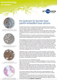 For an optimal stainning result...
Protocol for carrying out immunohistochemical labeling on floating sections
 Find here the protocol for performing immunohistochemical labeling on floating sections.
Sequential double stain protocol for immunohistochemistry
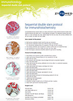
Double staining in immunohistochemistry for pathology laboratories can be easily achieved using the "Zytomed Systems Double Stain Polymer Detection Kit (POLDS-006)...
Microglie et maladie d'Alzheimer
La microglie est une population de macrophages située dans le système nerveux central (SNC), dont le premier rôle est la défense immunitaire du SNC. Des études ont montré que ces cellules pouvaient être impliquées dans le développement de la maladie d’Alzheimer.
SARS-CoV-2
Le nouveau coronavirus (SARS-CoV-2) a été découvert début janvier dans la ville de Wuhan (Chine) et s'est propagé dans le monde entier en quelques mois. La recherche de traitement efficace passe par l'étude du virus et DiagOmics se mobilise aux côtés de la communauté scientifique pour le combattre.
Range of Flow Cytometry reagents.
Find here our range of antibodies and reagents for flow cytometry.
Antibodies specific to collagen fibers
Find here all our antibodies directed against collagen fibers.
Compartmentalized boxes for your Western Blots
Our new compartmentalized boxes for staining your Western Blot gels or membranes are finally available !!!
Increasing Sensitivity ECL Range - WesternBright ECL
HRP sensitive substrate for Western blots
More efficient and faster transfer buffer
FLASHBLOT buffer improves protein transfer on western blot membranes..
Spectra Dye Antibody Labeling Kits
Spectra Dye Antibody Labeling Kits contain everything you need to couple antibodies ...
Western Blot Strip-It Buffer
Advansta Western Blot Strip-It buffer uniformly removes antibodies from Western blot membranes ...
TintoRetriever Pressure Cooker
The BioSB TintoRetriever pressure cooker rigorously controls the temperature and duration of antigenic or nucleic acid pre-treatment of tissues fixed with formalin and included in paraffin ...
 Download
ELISA Protocol
 Enzyme-Linked Immunosorbent Assays (ELISA tests) are used to measure an unknown concentration of antigen or antibody.
Antibody Stability - Antibody Diluent
The stability of diluted antibodies depends on several factors.
Pre-treatment for formalin-fixed paraffin-embedded tissue sections
For an optimal stainning result...
Protocol for carrying out immunohistochemical labeling on floating sections
Find here the protocol for performing immunohistochemical labeling on floating sections.
Storage tips for Advansta WB products
Find here some tips for storing products from the Advansta Western Blot range.
Sequential double stain protocol for immunohistochemistry
Double staining in immunohistochemistry for pathology laboratories can be easily achieved using the "Zytomed Systems Double Stain Polymer Detection Kit (POLDS-006)...